Basic Science Imaging Platform
Center for Anatomy and Cell BiologyEpiscopic 3D-Imaging
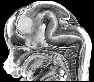
 Episcopic 3D imaging creates high-resolution digital volume data from biopsy material and sacrificed embryos. Platform members employ the Epi-3D, EFIC, and HREM methods for answering their scientific questions. Especially the Weninger Lab was significantly involved in developing these methods.
Brief technical description
Specimens are embedded in special resin or wax mixtures and sectioned on a microtome. During sectioning digital images of the subsequently exposed block surface are captured. The resulting stacks of approximately 500 to 3 000 inherently aligned and undistorted digital images can be immediately three-dimensionally (3D) visualised and analysed with all modern 3D visualisation software packages. Typical volume data, having a voxel size of 1µm x 1µm x 2µm and covering a volume of approximately 2mm x 3mm x 4mm are produced within 6 hours data generation time. (WJ Weninger, Dec. 2011)
Related Scientific Publications (Weninger Lab)
Split in topics: HREM-Embryos; HREM-Organic Material; HREM-Biopsies